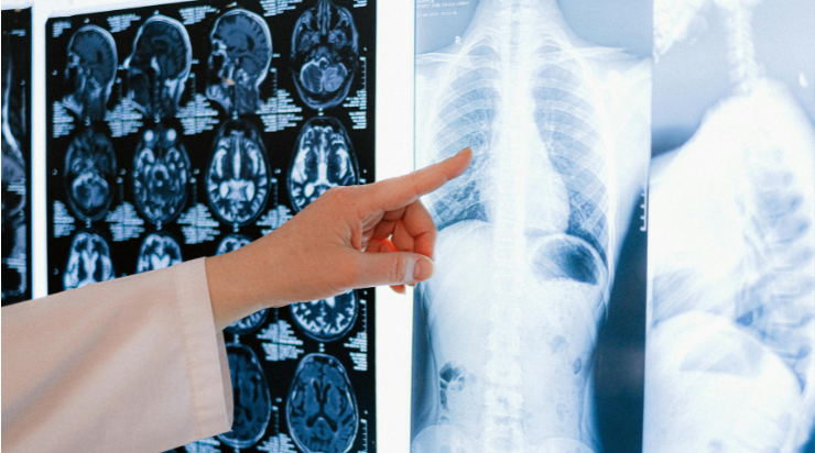
How AI is Revolutionizing Radiology Safety
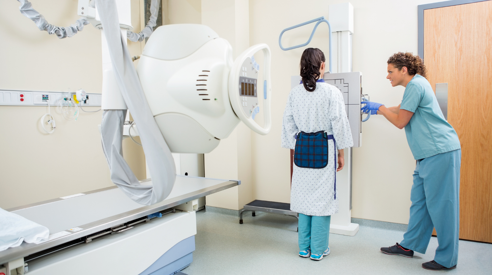
In the high-stakes world of radiology, every interpretation carries immense responsibility. With 3.1 billion diagnostic imaging studies performed worldwide each year, and chest X-rays accounting for 40% of that staggering volume, the pressure on radiologists is unrelenting. Yet despite decades of experience and technological advancement, diagnostic errors persist at 3–5% across all studies, translating to approximately 40 million missed or delayed diagnoses annually. These aren’t the result of incompetence or negligence, instead, 84% of these errors are perception failures, where abnormalities were objectively visible but simply overlooked by even the most skilled eyes.
Perception errors arise from fundamental human limitations that no amount of training can completely eliminate. Visual fatigue sets in predictably after interpreting 20–30 consecutive studies, reducing detection sensitivity by 15–20%. Cognitive biases like “satisfaction of search,” where identifying one abnormality causes radiologists to subconsciously terminate their visual search, influence 40% of multi-pathology misses. Anatomically complex regions, such as lung nodules obscured by overlying ribs or subtle pneumothoraces hidden behind scapulae, account for 62% of perception failures. Peripheral zones like costophrenic angles receive disproportionately less attention, leading to 25% of misses occurring outside primary focus areas.
The clinical consequences extend far beyond individual case errors. A landmark study of 1,000 delayed diagnoses revealed an average lag of 251 days between the initial miss and correct identification. Alarmingly, one-third of these errors never self-corrected, even when patients returned with objectively worsening symptoms. Lung cancer represented 28% of misses, pulmonary embolism 15%, aortic aneurysms 12%, and fractures 10%. In emergency settings, the stakes escalate dramatically, a missed pneumothorax carries 20% mortality if undetected, while overlooked pneumonia in elderly patients doubles 30-day mortality rates.
Workload emerges as the single most modifiable error multiplier. Comprehensive analysis of 2.9 million teleradiology interpretations established clear thresholds, shifts exceeding 10 hours demonstrated 67% higher error rates. High-volume shifts (67–90 studies) produced errors 226% higher than appropriately paced shifts (19–26 studies). Most concerning, radiologists interpreting at double their baseline speed experienced 270% higher error rates, a direct refutation of productivity incentives that prioritize volume over thoughtful analysis.
Burnout exacerbates this vicious cycle. Fifty-three percent of radiologists report clinical burnout, with 36% regularly working night shifts despite well-documented 50% higher diagnostic errors during circadian low points. Daily time pressure affects 42%, while 70% report work-family interference and 73% believe their job negatively impacts physical health. Teaching suffers too, radiologists in training programs spend 19% less time educating despite rising volumes, perpetuating workforce shortages.
AI second readers: Clinical evidence that delivers
Enter artificial intelligence as the tireless second reader, a clinical teammate that analyzes every study independently while preserving the radiologist’s authoritative judgment. Recent meta-analyses provide irrefutable evidence of impact. For pneumonia detection across 20 studies involving 15,000+ patients, radiologists alone achieved 85% sensitivity. Paired with AI second readers, sensitivity climbed to 95% with preserved specificity. Lung nodule detection, particularly for challenging ≤10 mm lesions, improved by 9–10 percentage points in screening programs analyzing over 25,000 chest X-rays.
Efficiency gains compound these clinical benefits. Prospective studies document a 36% reduction in reading time alongside a 15.94% AUC improvement, 11.44% sensitivity gains, and 2.95% specificity enhancement. Critically, AI captured 89% of radiologist errors that retrospective quality assurance would otherwise miss, preventing patient harm proactively.
Multimodal AI platforms further elevate performance by integrating X-ray, CT, and MRI data simultaneously. Rather than siloed single-modality analysis, multimodal systems cross-reference findings, detect inconsistencies that might represent artifacts or true pathology, and generate synthesized confidence scores that dramatically improve decision-making in complex cardiopulmonary cases where single studies prove inconclusive.
Real-world deployment: Hospitals leading the way.
Academic medical centers processing 1 million+ studies annually report transformative outcomes. One system implementing AI quality assurance caught 89% more errors before reaching patients, preventing an estimated 4–6 serious adverse events yearly while eliminating $1.2 million in potential liability exposure. European teleradiology networks achieved 37% reading time reduction, 12% detection improvement, and 28% radiologist satisfaction gains, sustained results, not pilot promises.
Streamlined clinical workflow
Implementation proves remarkably intuitive. Radiologists perform their initial read using clinical expertise while AI analyzes the identical study set in parallel, surfacing only high-confidence misses accompanied by visual evidence overlays and probability scores. Clinicians can confirm concordant findings, override discordant ones, or dismiss low-relevance alerts, all within existing workflow patterns. No notification deluge disrupts focus. Comprehensive audit trails document clinical oversight, strengthening medicolegal defense while enabling continuous performance improvement.
Compelling economic rationale
The business case is equally strong. A single missed malignancy incurs $500,000–$1 million in liability plus escalated treatment costs. AI second readers capturing merely 10% of misses prevent multiple claims annually. Enterprise implementation recoups investment within 3–6 months through liability avoidance, reduced re-read volume, and efficiency gains equivalent to hiring additional FTEs, without recruitment costs.
Radiologist perspectives: Voices from the trenches
Surveys of 1,200+ practicing radiologists reveal broad acceptance. Seventy-three percent report reduced backlog pressure, 72% note improved timeliness of critical diagnoses, 68% experience less cognitive fatigue, and 91% view AI as genuine augmentation rather than a replacement threat. This consensus emerges from clinicians experiencing sustained deployment, not theoretical promise.
Practical 30-day implementation roadmap
Week 1: Establish confidence through frictionless piloting. Deploy across 10% volume with zero workflow disruption. Capture baseline metrics for error rates, turnaround times, and satisfaction.
Weeks 2–3: Focus on enablement. Conduct concise one-hour training emphasizing override protocols and evidence interpretation. Scale to 50% volume while gathering daily clinician feedback to refine highlighting thresholds.
Week 4: Achieve full confidence. Complete 100% rollout supported by real-time performance dashboards tracking key metrics.
Month 2: Optimize for sustainability. Fine-tune confidence cutoffs based on departmental patterns, conduct formal satisfaction surveys, and quantify ROI through liability modeling and efficiency benchmarking.
Night shifts demonstrate the fastest ROI. AI eliminates the 50% elevated error risk from circadian fatigue, ensuring morning teams receive reliable handoffs.
Addressing legitimate clinical concerns
Liability exposure remains a primary worry. Comprehensive audit trails document adherence to augmented care standards increasingly recognized as best practice by medicolegal experts.
Alert fatigue plagues many systems. Intelligent filtering surfaces only the top 5% confidence misses with contextual visual evidence, preserving attentional resources.
Over-reliance risks vigilance erosion. Observed override rates of 15–20% confirm preserved independent judgment, while AI learns from clinician corrections to refine future performance.
Night shift transformation
Where human performance dips 50% due to circadian disruption, AI maintains unwavering consistency. Second-reader validation ensures off-hours interpretations match daytime quality, dramatically improving patient safety during vulnerable periods while reducing morning discrepancy reviews.
Evolutionary horizon
Current second readers are evolving toward predictive capabilities. Risk stratification will prioritize the highest-adverse-outcome potential, while automated follow-up recommendations optimize cascade imaging. Population-level analytics identify departmental miss patterns for targeted education.
Redefining clinical standards
Three to five percent error rates have endured as “acceptable” for 75 years. AI second readers render this tolerance obsolete. Solo interpretation without quality assurance validation will soon parallel reading film without digital PACS, archaic by modern standards.
Health systems adopting multimodal AI establish leadership in clinical safety. Radiologists leveraging second readers deliver near-perfect actionable finding detection while experiencing reduced burnout and enhanced professional satisfaction.
Even if we don’t talk about specific imaging modalities, radiology talent in general remains critically scarce across the globe.
Immediate action items
Schedule a 15-minute platform demonstration targeting your highest-volume rotation,
Launch a 10% volume pilot capturing baseline metrics,
Quantify 30-day outcomes across error capture, efficiency, and satisfaction,
Use evidence-based benchmarks to guide scaled adoption.
Futuuri transforms radiology from error-tolerant to error-intolerant. Fewer misses, less clinician stress, transformative patient outcomes.